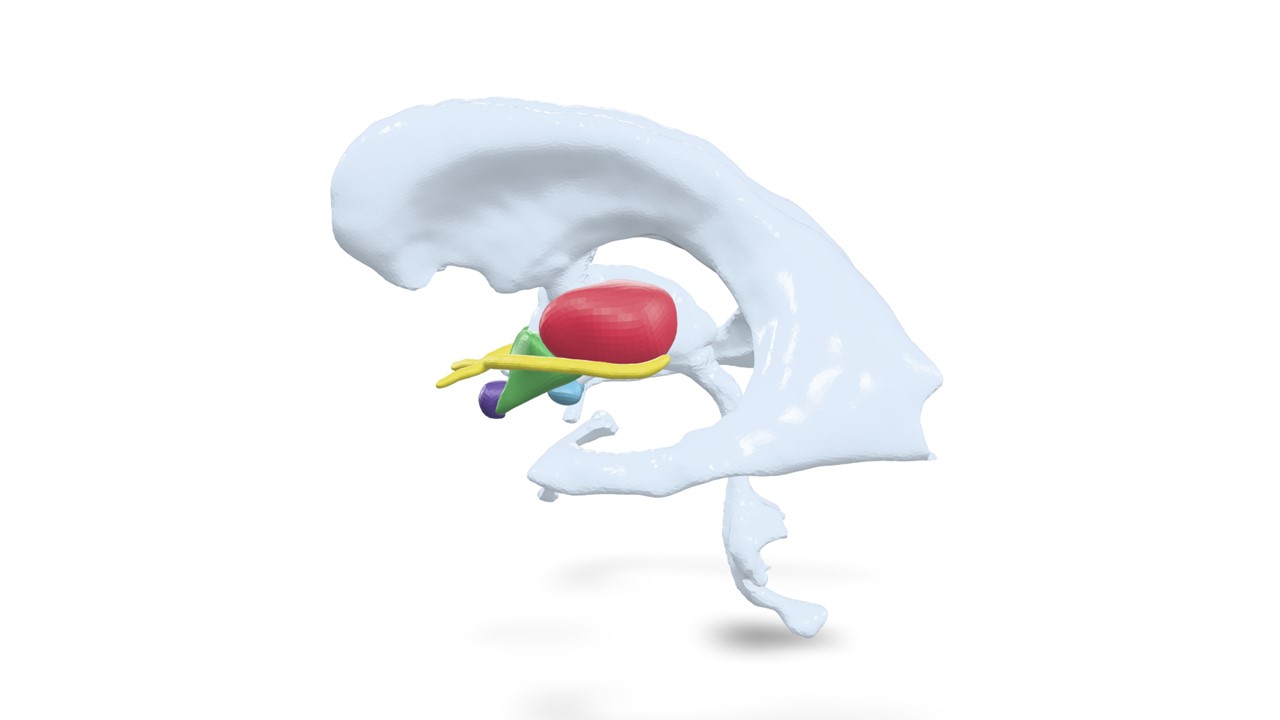
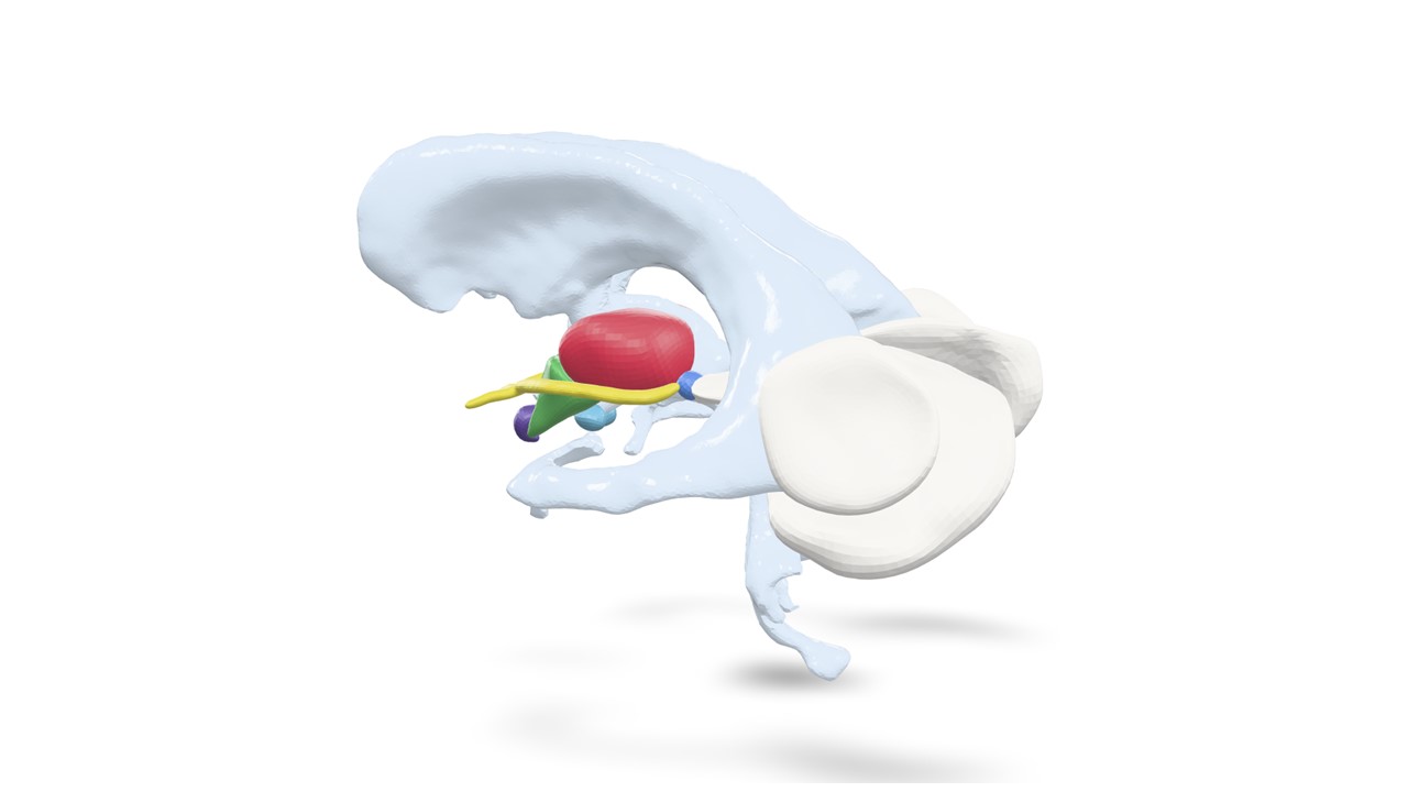
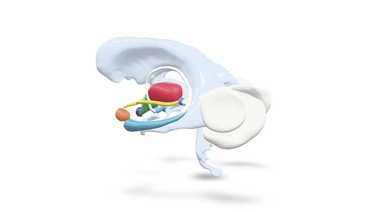
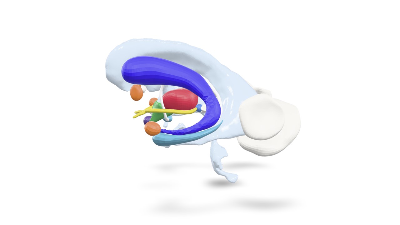
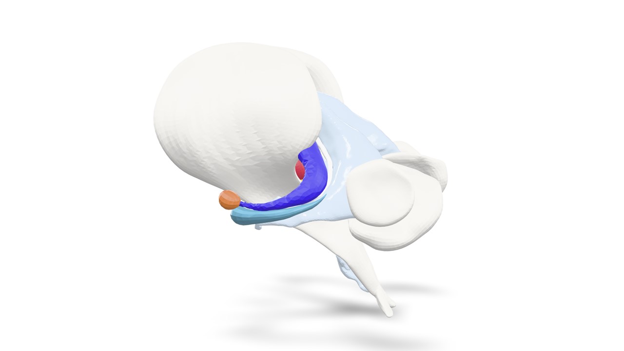
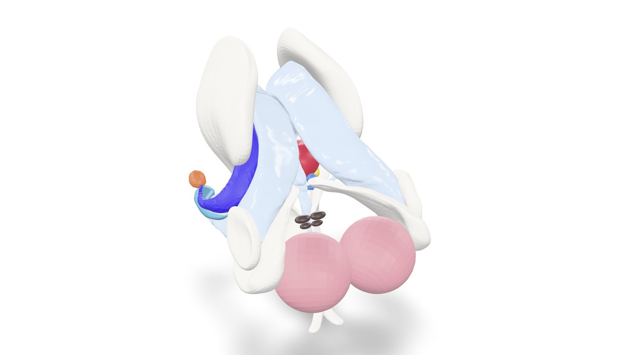
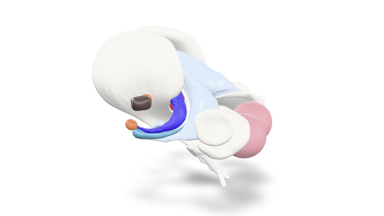
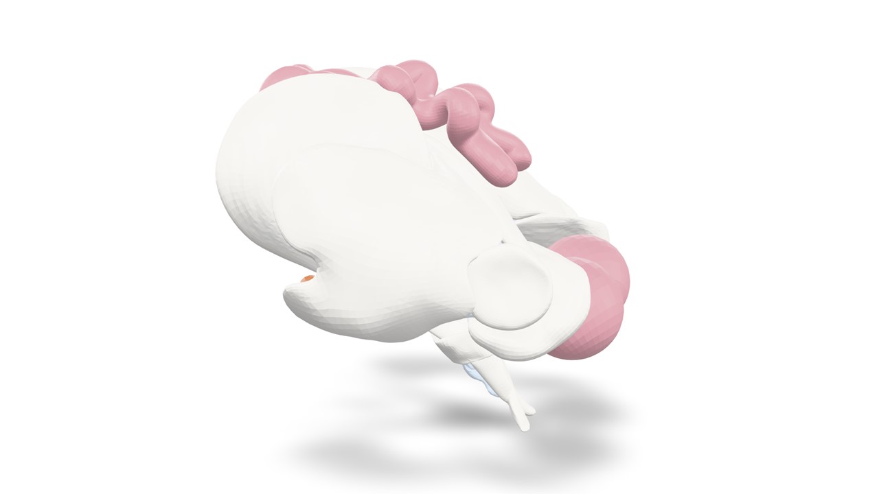
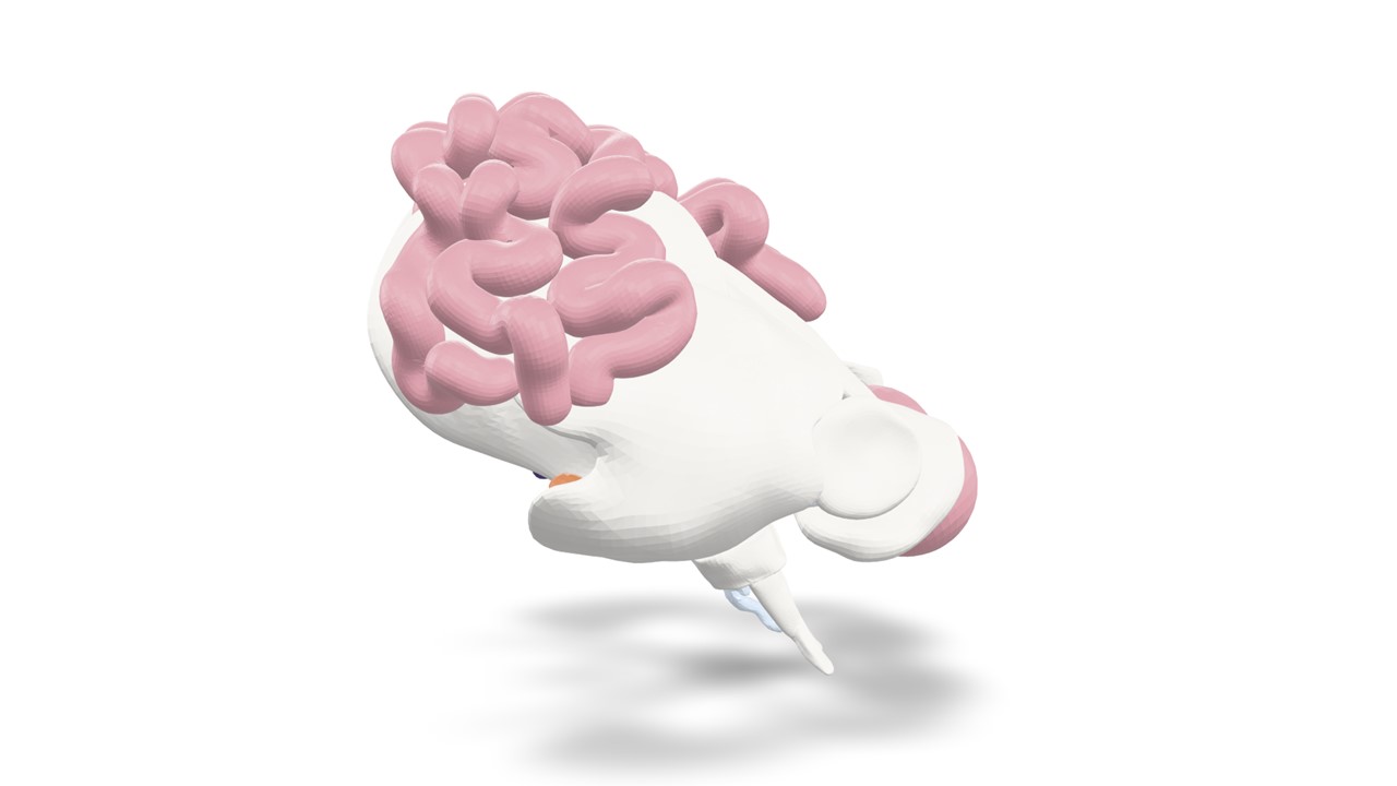
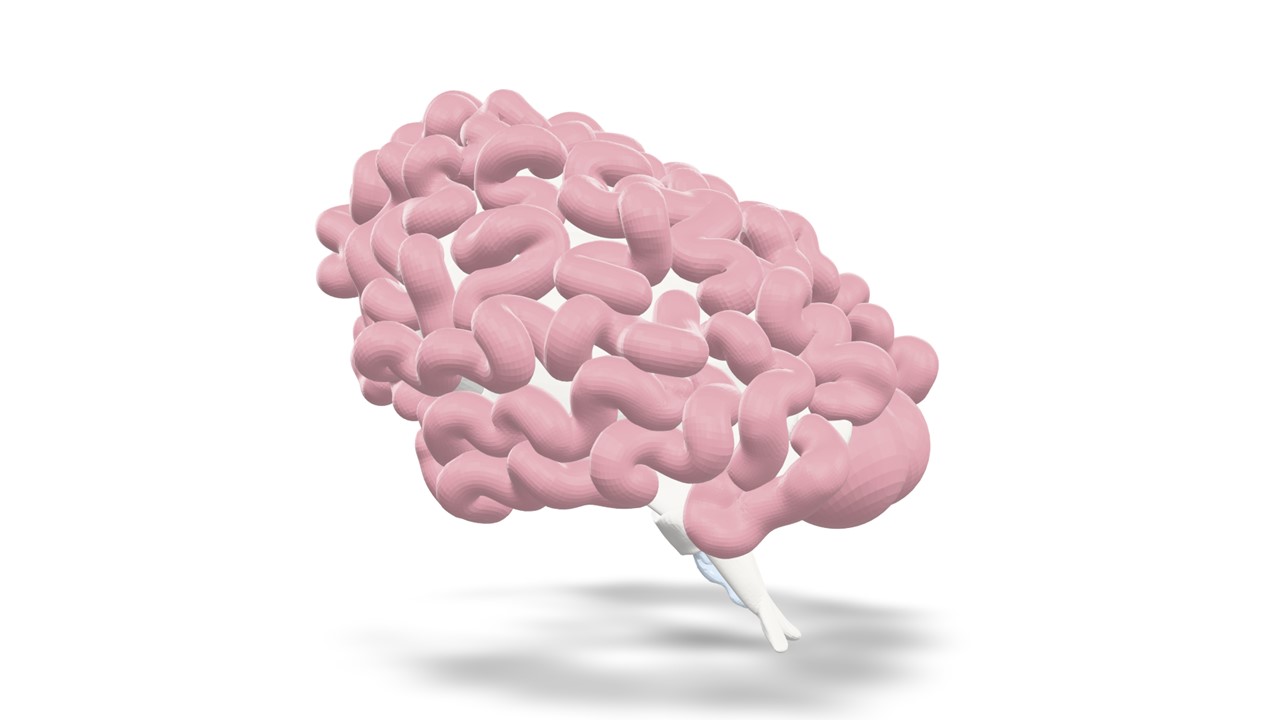
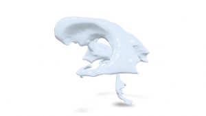
The human ventricular system is a good reference point for accurately reconstructing neuroanatomical structures. You can model neuroanatomical structures around it in lateral, dorsal, and ventral directions. Ventricles are ‘cavities’ in the brain tissue that are filled with cerebrospinal fluid (‘in vivo’). We use this ventricular model, in which the ‘cavities’ are filled with plastic, to attach the neuroanatomical structures. First, identify the following structures and areas in the ventricular model:
- Both lateral ventricles;
- Both frontal horns of the lateral ventricles;
- Both temporal horns of the lateral ventricles;
- Both occipital horns of the lateral ventricles;
- Both foramina of Monro (interventricular foramina);
- 3rd ventricle;
- Cerebral aqueduct of Sylvius;
- 4th ventricle;
- Foramina of Luschka;
Foramen of Magendie;
Central canal.

00 Ventricular system

01 Thalamus, Hypothalamus, Pituitary Gland, Optic Chiasm, Mammillary Bodies

02 Optic Radiation

03 Limbic System

04 Reward System

05 Corticospinal Tracts

06 Cerebellum

07 Basal Ganglia

08 Connections: Corpus Callosum, Cingulate Gyrus, and White Matter Networks

09 Structure of the Cerebrum, Starting with Three Important Gyri

10 Structure of the Cerebrum, Frontal, Parietal, and Occipital Lobes
